Figure 5.

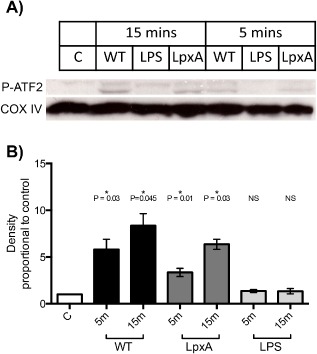
ATF2 phosphorylation is stimulated by WT and lpxA‐ bacteria, but not LPS.
A. Representative Western blot showing ATF2 phosphorylation in HUVEC after 15 min stimulation by 108 cfu ml−1 fixed WT or lpxA‐ bacteria, or 10 ng ml−1 LPS. COX IV antibody was used as a loading control.
B. Summary of mean ATF2 phosphorylation presented as fold increase in band density above control ± SEM, n = 3.
