
Figure 7.
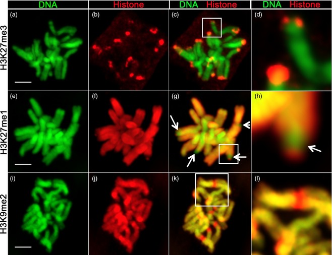
Immunolocalization of histone marks on barley mitotic chromosomes.
All chromosome spreads are from mitotic barley root tip cells: top row, histone marks H3K27me3 (a–d); middle row, H3K27me1 (e–h); bottom row, H3K9me2 (i–l). For each histone modification, DNA (green, Hoechst stain), histone mark (red), merge of DNA/histone mark and close‐up of the boxed region are shown. Bars = 5 μm.