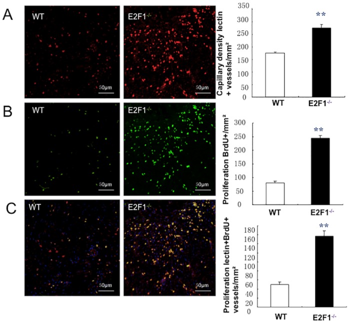
Fig 3. E2F1-deficiency enhances vessel growth in the border area of the skin wound.

At day 7 post-surgery, Lectin was i.v. injected 10 min before euthanasia to identify vasculature; subsequent analyses were performed in sections of wound skin and quantified at the border zone of wounds. (A) Functional vessels were identified by staining with anti-lectin antibodies (red) (left and middle panels, scale bar = 50mm) and quantified (right panel). (B) Proliferation cells were identified by staining for BrdU (green) (left and middle panels) and quantified (right panel). (C) Proliferating ECs (yellow) were identified by co-staining for BrdU (green) and lectin (red) (left and middle panels, scale bar = 50 mm) and quantified as the number of lectin+BrdU+ vessels per mm2 (right panel); nuclei were stained with DAPI (blue). **p<0.01; n = 10 per group.
