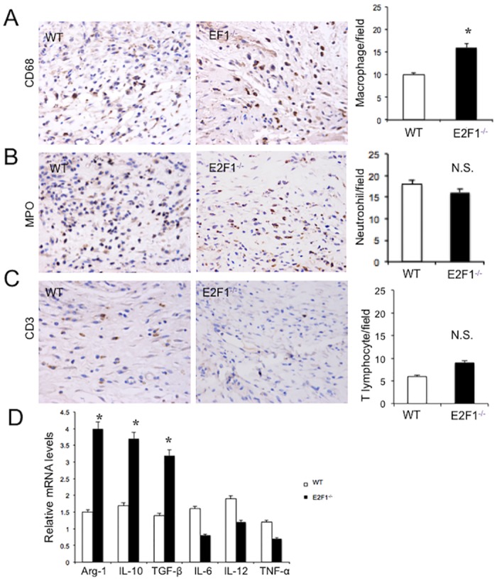
Fig 4. More infiltrating macrophages are found in the wound of E2F1–/– mice than in WT mice.

At day 7 post-surgery, the wound skin tissues were analyzed immunochemically and by qRT-PCR. Shown are representative staining (left and middle panels) (original magnification, X400; 0.06 mm2/field) and quantifications (right panels) of the cells stained positive for CD68 (A), MPO (B) and CD3 (C). (D) The expression levels of M1 and M2 macrophage marker genes in the wounded skin tissues of WT and E2F1–/– mice were analyzed by qRT-PCR. **p<0.01. n.s., not significant; n = 10 per group.
