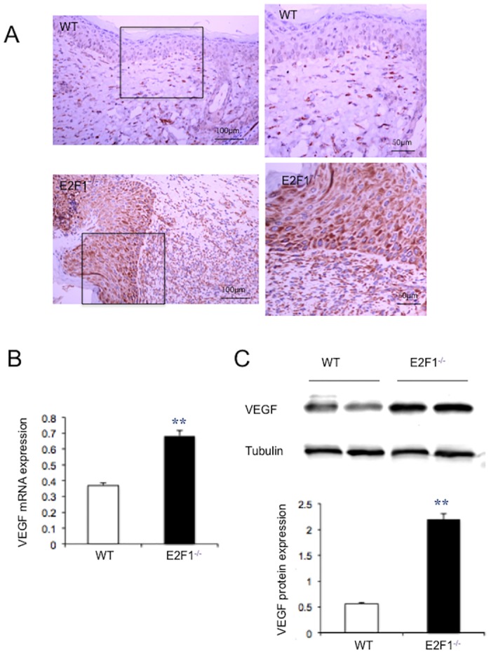
Fig 5. VEGF-A level is significantly higher in the wound of E2F1–/– mice than in WT mice.

Wound tissues were isolated at day 7 post-surgery. (A) Immumohistochemical staining of VEGF (brown), 20X original magnification (left panels), the inlets were enlarged (right panels). (B) VEGF mRNA expression was analyzed by qRT-PCR and normalized to the level of GAPDH. (C) Representatives of Western blotting (upper panel) and quantification of VEGF protein levels (lower panel). Protein levels were quantified densitometrically and normalized to tubulin levels. **p<0.01; n = 5.
