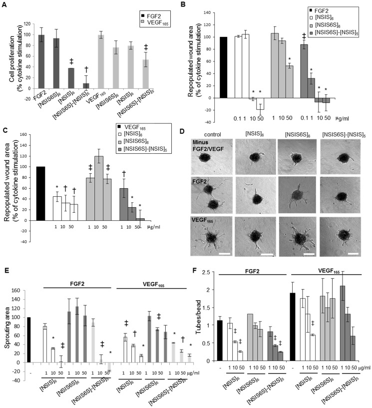
Fig 2. In vitro inhibitory potential of site-selectively 6-O-sulfated dodecasaccharides.
A, dodecasaccharides were tested for effects on FGF2- and VEGF165-induced HUVEC proliferation. The treatments were performed for 5 days. Stimulation of proliferation in response to FGF2 (20 ng/ml) and VEGF165 (20 ng/ml) is expressed as 100%. Dodecasaccharides were used at 50 μg/ml concentration. The mean ± SEM (n = 3) is shown. †, P < 0.01; ‡, P < 0.05. B to C, inhibition of HUVEC FGF2- and VEGF165-induced migration by dodecasaccharides was tested in wound healing assay. Wounds were created in confluent monolayers of serum-starved HUVEC and FGF2 (B) or VEGF165 (C) was added to promote cell migration into the wounds. Dodecasaccharides were used at a range of concentrations starting from 0.1 μg/ml for FGF2 and 1 μg/ml for VEGF165 with the highest concentration being 50 μg/ml in all assays. The wound area was measured at the beginning and 24 hours after the treatment. The area repopulated after 24 hours in response to growth factor alone is expressed as 100% (control). The effect of dodecasaccharides is expressed as a percentage of repopulated area induced by a growth factor alone. All experiments were performed three times in triplicates. The mean ± SEM (n = 3) is shown. *, P < 0.001; †, P < 0.01; ‡, P < 0.05. D, HUVEC spheroids were embedded in fibrin gels that were overlaid with either EBM-2 media lacking FGF2 and VEGF165, EBM-2 media supplemented with FGF2 (5 ng/ml) or VEGF165 (2.5 ng/ml) and EBM-2 media supplemented with FGF2 or VEGF165 and dodecasaccharides (50 μg/ml). The treatment was performed for 24 hours. Scale bars represent 200 μm. E, sprouting area in each spheroid was evaluated using Metamorph software where the area of outgrowing sprouts was derived by subtracting the area of a spheroid without sprouts from the total area of a spheroid. The increase of sprouting area after stimulation with FGF2 or VEGF165 is expressed as 100% (control). The ability of oligosaccharides to reduce FGF2 and VEGF165-induced endothelial cell sprouting is expressed as a percentage of control. 20–30 spheroids were analysed per each experiment and three independent experiments were performed. The values are shown as mean ± SEM (n = 3). *, P < 0.001; †, P < 0.01; ‡, P < 0.05. F, the effect of oligosaccharides on endothelial tube formation was evaluated in three-dimensional fibrin gel bead assay. FGF2 and VEGF165 were used at 5 ng/ml concentration. No endothelial tubes developed in the absence of FGF2 and VEGF165. Dosing was performed for 5 days. The ratio of average number of endothelial tubes per bead is shown. Three independent experiments were performed each in triplicate. The values are expressed as mean ± SEM (n = 3). *, P < 0.001; †, P < 0.01; ‡, P < 0.05.

