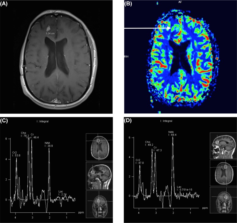
FIGURE 1.
A 31-year-old male patient with prior resection of a right frontal glioblastoma multiforme presented later with a 1-cm enhancing nodule concerning for recurrence compared with pseudoprogression. (A) Magnetic resonance image of the new nodule. (B) Map of relative cerebral blood volume demonstrates increased flow in the nodule (arrow). (C) Spectroscopic imaging demonstrates an elevated choline peak compared with (D) the normal contralateral brain parenchyma in the same patient.

