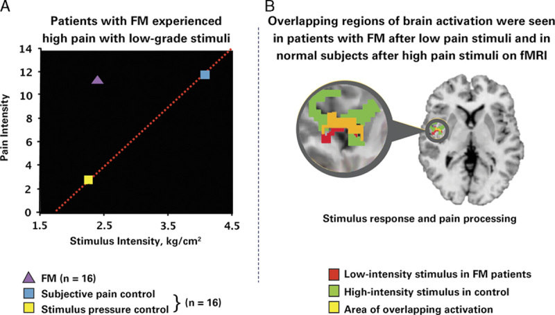
FIGURE 3.

Objective evidence shows augmented pain sensitivity in individuals with fibromyalgia (FM) versus matched controls.27 A, The graph depicts mean pain ratings plotted against stimulus intensity. In FM patients, a low stimulus pressure (2.4 kg/cm2) produced a much higher pain level (mean±SD, 11.30±0.90) than in controls. However, in control subjects, a much higher stimulus pressure elicited a pain response similar to that in FM patients. B, The scan is a functional magnetic resonance image (fMRI) of the brain of a patient with FM from the same study. The imaging study demonstrated that, in patients with FM, pain processing areas of the brain are activated at a much lower level of stimulus than in control subjects. There is overlap (as indicated by the yellow area on the fMRI) between the areas activated with a low-intensity stimulus in FM patients (red area) and a high-intensity stimulus in control subjects (green area). In other words, the overlap between brain activation in FM patients receiving a low stimulus pressure and controls receiving almost twice as much pressure (ie, the amount required to cause the same amount of pain) suggests a mechanism involving central amplification of pain in the patients with FM. Because regions of brain activation in FM patients and healthy controls overlap, the pain experienced by both sets of subjects is real. Objective evidence such as this would be a valuable component of the educational framework proposed in this publication. Gracely et al.27 Reprinted with permission from John Wiley and Sons. Copyright [John Wiley and Sons, New York, NY]. All permission requests for this image should be made to the copyright holder.
