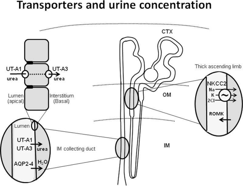
Figure 1.

Diagram of transporters involved in urine concentration. In the center is a diagram of the nephron with cortex (CTX), outer medulla (OM) and inner medulla (IM) marked. Oval enlargements show locations of the sodium potassium 2 chloride cotransporter (NKCC2) and the renal outer medullary potassium channel (ROMK) in the thick ascending limb, aquaporins 2-4 (AQP2-4) in the inner medullary collecting duct (IMCD) and urea transporters (UT-A1 and UT-A3) in the IMCD. Top left enlargement shows IMCD cells with UT-A1 located apically and UT-A3 located basolaterally.
