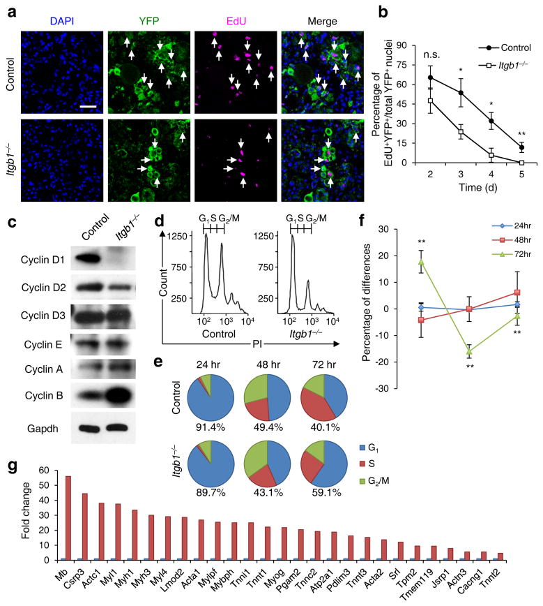
Figure 2.
Young Itgb1−/− SCs are defective in maintaining proliferation and prone to differentiation. (a) EdU incorporation of YFP+ control and mutant cells in muscle sections 3 d after injury; Scale bar, 50 μm. (b) Quantification of EdU+YFP+/YFP+ on d 2 – 5 daily; data are expressed as mean ± s.d., n = 3 animals per d; ten sections per sample; Student’s t-test: *P < 0.05, **P < 0.01, and n.s., non-significant. (c) Western blot of FACS isolated control and Itgb1−/− YFP+ SCs cultured for 72 h, using antibodies to proteins indicated. (d–f) FACS-aided cell cycle analyses using DNA content (stained by PI) of control and mutant SCs at 24, 48, and 72 h: (d) PI profiles at 72 h, (e) pie charts summarize cell fractions in G1, S, and G2/M, and (f) percentage deviation plot of mutant vs. control cells in cell cycle phases at stipulated time points; ModFit LT V2.2.11 was used to analyze percentages of each phase of the cell cycle. All data were determined to have “good” RCS, measurement of fit. (g) Fold changes of muscle differentiation genes upregulated in Itgb1−/− SCs compared to control SCs at 72 h. All listed genes display “yes” significance (q < 0.05) by Cuffdiff2, Genes were included only if one (control or Itgb1−/−) had FPKM ≥5 to control for elevated fold changes of genes with minimal expression.

