Figure 1.

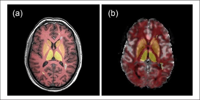
A representative slice of (a) T1-weighted MRI scan and (b) map of cerebral blood flow derived using the bookend technique with gray matter, white matter, basal ganglia, thalamus, and white matter lesion tissue regions of interest overlaid.

A representative slice of (a) T1-weighted MRI scan and (b) map of cerebral blood flow derived using the bookend technique with gray matter, white matter, basal ganglia, thalamus, and white matter lesion tissue regions of interest overlaid.