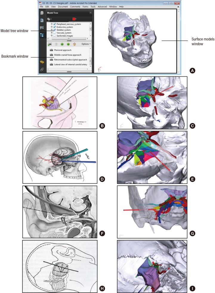
Fig. 3.
In the PDF file, windows showing each 3D model and bookmarks of clinical trials. Images from left column are derived from references: B (22), D (18), F (20), and H (21). In the model tree, bookmark, and surface models windows of the PDF file, user can select 3D models of each structure, sectioned images, and clinical trial (A). Regarding clinical approaches, textbook figures (B, D, F, H) and correspondence with 3D models of this study (C, E, G, I) are shown. Interhemispheric approach through the superoposterior and superior triangles (B, C), pterional approach through the middle, inferior, anteromedial, and anterolateral triangles (D, E), middle cranial fossa approach through the posterolateral and posteromedial triangles (F, G), and retromastoid suboccipital approach through the lateral and medial triangles (E) can be operated virtually.

