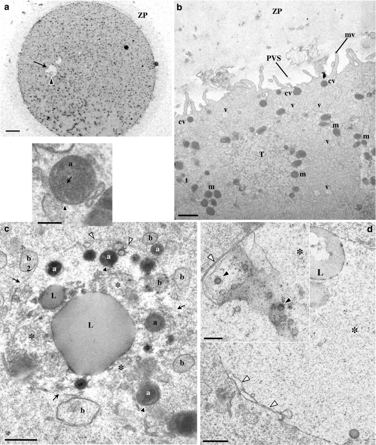
Fig. 3.

Ultrastructure of human mature oocyte with a small granular vacuole. a Semithin section. The inclusion appears as a round structure with a pale appearance (arrow), and at one pole, there is the presence of an aggregate of moderate dense material (black arrowhead). Note the zona pellucida (ZP). b–d Ultrathin sections. b At the oocyte cortex, cortical vesicles (cv), isolated smooth endoplasmic reticulum (SER) vesicles (v) and tubules (t), a SER tubular aggregate (T), and moderate dense mitochondria (m) can be observed. Note the perivitelline space (PVS) and microvilli (mv). c The granular vacuole presents regions delimited by a membrane (white arrowheads) and regions with membrane disruption (arrows). The interior of the granular vacuole presents a fine fibrillar appearance (asterisk). Within the granular vacuole, there are small dense vesicles (a) with a double membrane and cristae (black arrowheads; also see inset), small vesicles with fine fibrillar materials (b), small lipid droplets (L), and a very large lipid droplet (L). d Periphery of the granular vacuole (asterisk). Note regions with double membrane (white arrowheads). Similar structures to nuclear pores (black arrowheads) could also be observed (inset). a 20 μm. b 1 μm. c 0.5 μm. Inset in c 0.25 μm. d 0.5 μm. Inset in d 0.25 μm
