
Fig. 4.
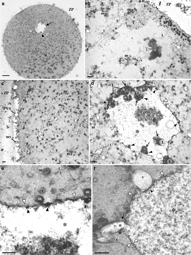
Ultrastructure of human mature oocyte with a medium-sized granular vacuole. a Semithin section. This vesicle appears as a round pale structure (arrow) with peripheral inner small aggregates of materials with moderate density (black arrowhead). Note de zona pellucida (ZP). b–f Ultrathin sections. b At the oocyte cortex, there are cortical vesicles (cv), smooth endoplasmic reticulum (SER) isolated vesicles (v) and tubules (t), a SER tubular aggregate (T), and moderate dense mitochondria (m). Note the metaphase II plate (white arrowhead). c Oocyte cortex and subcortex. Note the abundance of SER isolated tubules and mitochondria, small MV complexes, the perivitelline space (PVS), and microvilli (mv). d The granular vacuole presents regions delimited by a membrane (white arrowheads). At the periphery, there is inclusion of moderate dense lipid droplets (L) associated to dense materials (arrows). There is also inclusion of small round dense structures (black arrowheads). The interior of the granular vacuole presents a fine fibrillar appearance (asterisk) with a granulo-fibrillar dense region at the center (n). e Periphery of the granular vacuole. Note regions with double membrane (white arrowheads), the dense cover of the inner membrane, and the associated small round dense structures (black arrowheads). f Periphery of the granular vacuole. Note regions with double membrane (white arrowheads), a granular vacuole protrusion (arrow), and a granular vacuole indentation (black arrowhead) in association with a vesicle (v) containing floccular contents. a 20 μm. b–d 1 μm. e 0.5 μm. f 05 μm