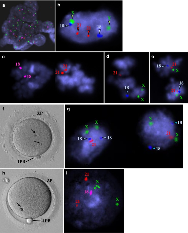
Fig. 5.

Images from epifluorescence microscopy using fluorescent in situ hybridization (FISH) analysis of human mature oocytes for detection of chromosomes X, 18, and 21 (a–e, g, i). Live human mature oocytes with small granular vacuoles observed at the inverted microscope (f, h). a Residual follicular cells were present in one case and showed a diploid constitution (XX,1818,2121). X (green), 18 (pink), 21 (red). b Polar body from one case showing a diploid constitution (XX,1818,2121). X (green), 18 (blue), 21 (red). c–e Metaphase II from three cases. c Diploid constitution due to probable replication associated with monosomy X (X,1818,2121). X (green), 18 (pink), 21 (red). d Haploid constitution with replication of chromosome X (XX,18,21). X (green), 18 (blue), 21 (red). e Diploid constitution due to probable replication (XX,1818,2121). X (green), 18 (blue), 21 (red). f Mature oocyte showing two small granular vacuoles. g Both small granular vacuoles showed a diploid constitution with monosomy 21 (XX,1818,21). X (green), 18 (blue), 21 (red). h Mature oocyte showing a small granular vacuole. i The small granular vacuole shows a diploid constitution with monosomy 18 (XX,18,2121). X (green), 18 (pink), 21 (red)
