FIGURE 7.
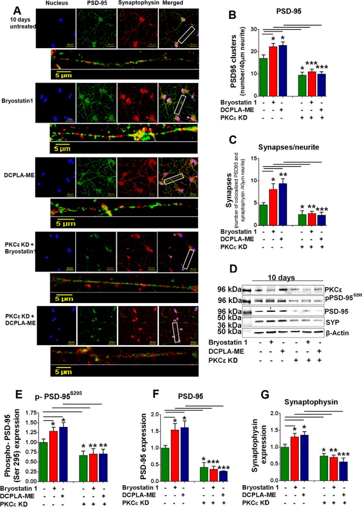
Loss of PKCϵ prevents synaptogenesis. A, confocal images of untreated and DCPLA-ME (100 nm)-, bryostatin 1 (0.27 nm)-, PKCϵ siRNA + DCPLA-ME (100 nm)-, and PKCϵ siRNA + bryostatin 1 (0.27 nm)-treated primary human neurons (10 days). Each condition is represented by five panels. Four square panels represent nucleus (blue), PSD-95 (green), synaptophysin (red), and merged image. The rectangular panel represents a magnified image of a 40-μm neurite. B, number of PSD 95 signal grains was measured along 40-μm neurite length (10 individual neurites from 4 independent slides). Bryostatin 1 and DCPLA-ME significantly increased the PSD-95 clusters per 40 μm neurite (F(2,9) = 4.5; ANOVA p < 0.05). C, synapses were quantified by the number of colocalized PSD-95 and synaptophysin signals. PKCϵ activation increased synapse number (F(2,9) = 6.1; ANOVA p < 0.05), and PKCϵ KO prevented the synaptogenic effect of PKC activators. D, immunoblot analysis of PKCϵ, p-PSD-95S295, PSD-95, and synaptophysin. PKCϵ knockdown (KD) reduced PKCϵ expression by 50% after 10 days in human neurons. E–G, at 10 days PKCϵ activation increased the expression of PSD-95 and synaptophysin significantly, but in PKCϵ KD cells their expressions were lower even after treatment with activators. Data are represented as the mean ± S.E. of at least three independent experiments (Student's t test. *, p < 0.05; **, p < 0.005; ***, p < 0.0005).

