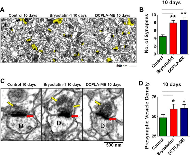
FIGURE 8.
PKCϵ activation induces synaptogenesis in hippocampal slices. A, electron microscopy of the hippocampal CA1 area from adult organotypic brain slices treated with vehicle (Control), DCPLA-ME (100 nm), and bryostatin 1 (0.27 nm) for 10 days. Dendritic spines showing synapse are highlighted in yellow. B, PKCϵ activation by bryostatin 1 and DCPLA-ME increased synapse number in adult organotypic brain slices (F(2,6) = 11.9; ANOVA p < 0.01). C, electron micrograph showing increased presynaptic vesicle density in PKC activator treated slices. The gray level of presynaptic vesicle stack from six to seven presynaptic boutons was measured from three different hippocampal slices. D represents dendritic spine, the red arrow marks synapse, and yellow marks presynaptic vesicles. D, bryostatin 1 and DCPLA-ME significantly induced the presynaptic vesicle density at 10 days. Data are represented as the mean ± S.E. of at least three independent experiments (Student's t test. *, p < 0.05; **, p < 0.005).

