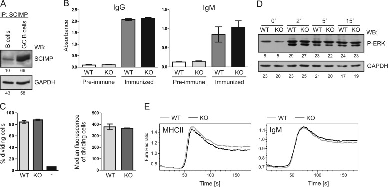
FIGURE 2.
Normal responses of Scimp−/− B cells. A, sorted naïve mouse B cells from control mouse or GL7+ germinal center (GC) B cells from sheep red blood cell-immunized mice were lysed in SDS-PAGE sample buffer and probed for SCIMP protein by immunoblotting. GAPDH staining served as a loading control. IP, immunoprecipitation; WB, Western blotting. B, sera from WT (n = 5) or Scimp−/− (KO, n = 6) mice immunized intradermally with ovalbumin in incomplete Freund adjuvant were collected, and immunoglobulins specific to ovalbumin were detected by ELISA. As a control, preimmune sera from the same mice were used. C, proliferation of OTII transgenic T cells labeled with CFSE was measured after 48 h co-culture with NP-ovalbumin-fed B1–8i transgenic B cells (WT or Scimp−/−) by flow cytometry. T cells cultured alone served as a negative control (−). Percentages of dividing cells and median CFSE fluorescence of cells that underwent at least one division are shown. D, ERK1/2 phosphorylation after MHCIIgp cross-linking in WT and Scimp−/− primary splenic B cells was analyzed by immunoblotting. E, splenocytes isolated from WT and Scimp−/− mice were stained with APC-conjugated anti-CD3 and anti-CD11b antibodies. The increase in calcium flux after MHCIIgp or IgM cross-linking was evaluated in B cells (gated as CD3− CD11b−) using a Fura Red calcium-sensitive probe.

