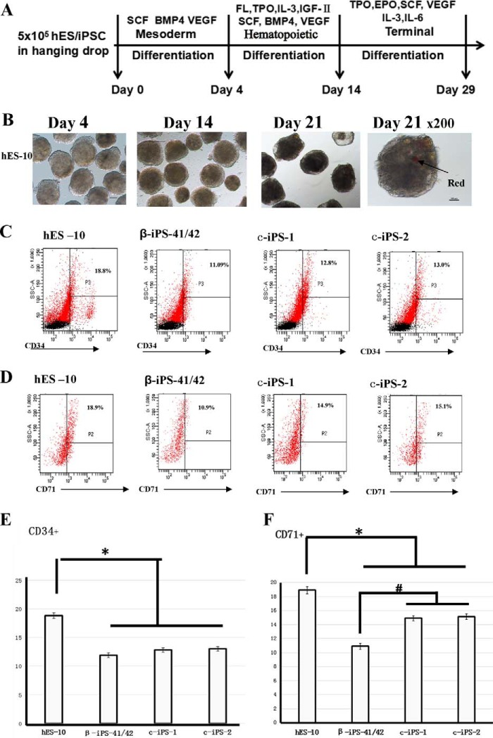
FIGURE 5.
Differentiation of β-Thal iPSCs and corrected iPSCs. A, schematic view of in vitro hematopoietic differentiation. Sequential stages are mesoderm induction, hematopoietic stem/progenitor cell emergence, and terminal differentiation. B, bright field images of β-iPS-41/42 cells differentiated for 4, 14, and 21 days. C and D, flow cytometry analysis of hES-10, β-iPS-41/42, c-iPS-1, and c-iPS-2 cell surface markers CD34 and CD71 after differentiation for 14 and 21 days. E and F, statistical results of flow cytometric analysis of hES-10, β-iPS-41/42, c-iPS-1, and c-iPS-2 cell surface markers CD34 and CD71 after differentiation for 14 and 21 days. Means ± S.D. (n = 3) are shown. *, p < 0.05 compared with the hES-10 group; #, p < 0.05 compared with the β-iPS-41/42 group. SCF, stem cell factor; EPO, erythropoietin; TPO, thrombopoietin; FL, Flt3 ligand; SSC, side scatter.

