FIGURE 9.
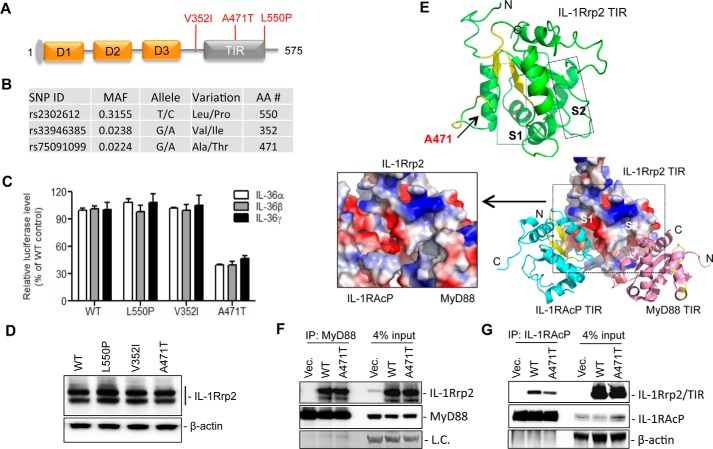
Effects of IL-1Rrp2 SNPs on signal transduction. A, locations of three non-synonymous SNPs in the IL-1Rrp2. B, properties of the IL-1Rrp2 SNPs. SNP data were derived from NCBI SNP database. C, comparisons of the effects of SNPs on IL-36R signaling in 293T cells. Cells were transfected with plasmids encoding WT IL-1Rrp2 or the SNPs. The cells were mock-treated or stimulated with three IL-36 ligands, respectively. The data were plotted relative to WT IL-1Rrp2. D, analysis of SNP expression in 293T cells. E, molecular modeling the TIR domain of IL-1Rrp2 and its interaction with TIR domain of MyD88 and IL-1RAcP. S1 and S2 denote the two grooves in the IL-1Rrp2 TIR domain that are modeled to interact, respectively, with the IL-1RAcP TIR and the MyD88 TIR. Ala-471 is located in the S1 groove. F, immunoprecipitation to examine the interaction between the FLAG-tagged WT or A471T IL-1Rrp2 TIR domain with MyD88. 293T cells were co-transfected with MyD88 along with either the WT IL-1Rrp2 or A471T for 24 h and then mock-treated or stimulated by IL-36γ for 5 h. The antibody used for IP was targeted against MyD88. MyD88 levels in the lysate and immunoprecipitated complex were detected as the control. G, immunoprecipitation assay to examine the interaction between the FLAG-tagged WT and A471T IL-1Rrp2 TIR domain with IL-1RAcP. Immunoprecipitation used the anti-IL-1RAcP antibody. The TIR domain of IL-1Rrp2 was detected with an anti-FLAG antibody. The data represent three independent experiments with consistent results.

