
FIGURE 7.
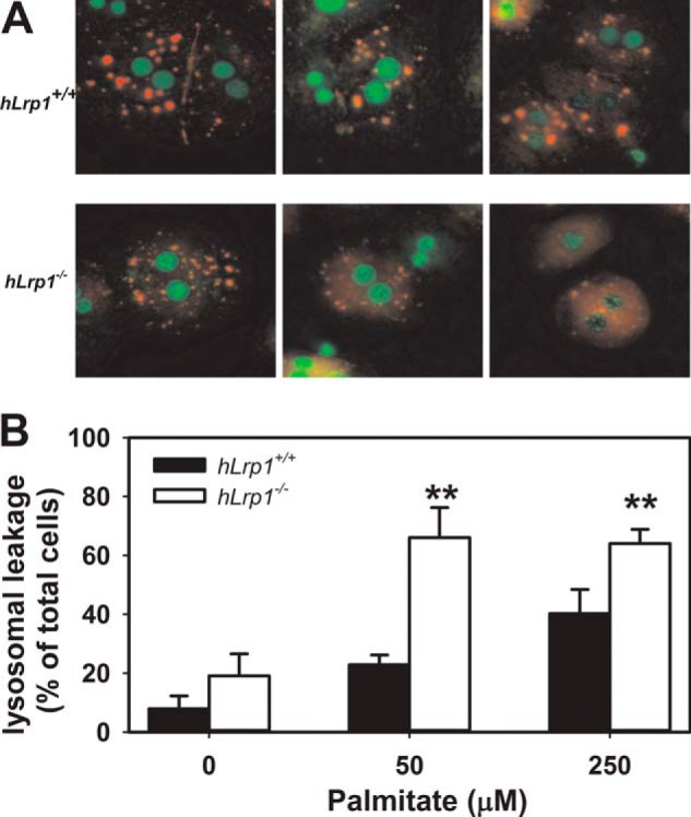
Palmitate-induced lysosomal permeability in hLrp1+/+ and hLrp1−/− hepatocytes. Primary hepatocytes obtained from hLrp1+/+ and hLrp1−/− mice plated on coverslips were treated with or without palmitate for 6 h and then stained with 4.64 μm acridine orange for 15 min. A, representative images with nuclei identified by green staining, intact lysosomes identified by punctate orange staining, and lysosomal leakage demonstrated with a diffuse orange staining pattern. B, mean ± S.E. (error bars) of data obtained by counting 200 cells in three separate experiments with duplicate cultures. **, significant difference from the hLrp1+/+ group at p < 0.01.