FIGURE 1.
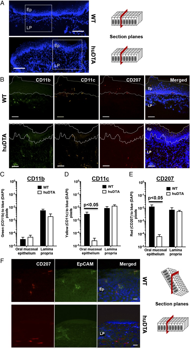
huLangerin-DTA mouse gingiva lack mucosal LCs at steady-state. Frozen maxillary gingival sections from C57BL/6J (wild-type [WT]) and huLangerin-DTA (huDTA) mice were stained with DAPI and probed with flurochrome-conjugated anti-mouse mAbs to detect CD207, CD11b, CD11c, and EpCAM. Sections were examined by confocal scanning laser microscopy. (A) DAPI signal on stained gingival sections taken at original magnification ×10 showing regions examined at higher magnification in (B). Scale bars, 100 μm. (B) Separate and merged images showing DAPI-, CD11b-, CD11c-, and CD207-specific staining of representative gingival sections (original magnification, ×40). Scale bars, 40 μm. Stratified squamous epithelium (Ep) and lamina propria (LP) regions are labeled and delineated by a dashed white line to facilitate the signal quantification summarized in (C)–(E). (C–E) Quantification of (C) CD11b-, (D) CD11c-, and (E) CD207-specific signals in Ep and LP regions as the ratio of green, yellow, or red to blue (DAPI) pixels, respectively. Pooled pixel ratios obtained from 11 separate sections from each mouse strain were compared using two-tailed Student t test and presented as mean ± SEM. (F) Representative separate and merged images showing DAPI-, CD207-, and EpCAM-specific staining in Ep or LP gingival regions taken from WT or huDTA mice, respectively (original magnification, ×40). Scale bars, 20 μm.

