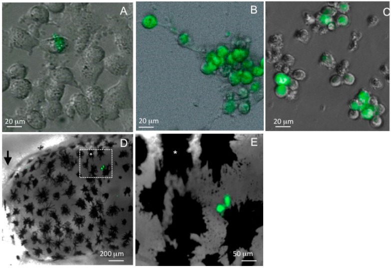
Figure 2.
Detection of FV3-GFP knock-in mutant expressing GFP reporter under the control of the immediate early 18K promoter during infection in vitro in mammalian cell lines and in vivo in X. laevis tadpoles. (A) BHK cells at 2 h post-infection at permissive (30 °C) temperature; (B) mouse BV2 macrophage-like microglial cells at 24 h post-infection at permissive (30 °C) temperature; (C) mouse sertoli macrophage TM4 at 24 h post-infection at non-permissive (37 °C) temperature; and (D,E) midbrain view of pre-metamorphic tadpole brain at 1 day post-infection at low (D) and higher (E) magnification. (*) Indicates the same melanophore in panel D and E. Images are composite of phase contrast and fluorescence for cells (A–C) and of bright field and fluorescence of the whole-mounted tadpole, taken under a Leica DMIRB inverted fluorescence microscope and Infinity 2 digital camera (objectives ×5/×10/×20; Zeiss). Digital images were analyzed and processed by ImageJ software.

