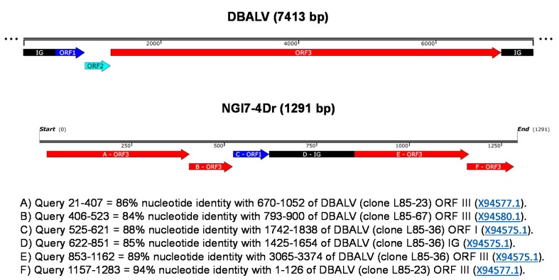
Figure 3.
Schematic representation of the rearranged ORF3 of badnavirus clone NGl7-4Dr (KX008602) amplified from D. rotundata TDr 1950B by RCA. The clone length is shown in the linear scale bar and the rearranged fragments are represented in panels A-F. A non-scaled linear view of the genome organization of DBALV is shown in the top panel. The intergenic region (IG) and open reading frames (ORFs) appear with the following colour codes (adapted from Umber et al. [30]): IG, black; ORF1, dark blue; ORF2, light blue; ORF3, red.

