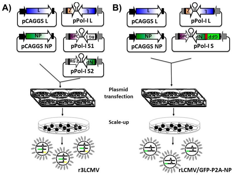
Figure 3.
Generation of recombinant replicating competent reporter-expressing arenavirus: Arenavirus rescues are performed in rodent (using the mouse Pol-I promoter) or in human (using the human Pol-I promoter) cells in six-well plates. Alternatively, arenavirus rescues can be performed in T7-expressing cells using plasmids driving the expression of the arenaviral S and L segments under the T7 promoter. (A) Generation of r3 arenaviruses: Cells are transiently co-transfected, using LPF2000, with the pCAGGS protein expression plasmids encoding the viral NP and polymerase L (required to initiate viral gene transcription and genome replication) together with the pPol-I vRNA expression plasmids for the viral L segment and the two (pPol-I S1 and pPol-I S2) viral S segments. In the pPol-I S1 plasmid, the viral NP is replaced by a reporter gene 1 (RG 1), and, in the pPol-I S2 plasmid, the reporter gene 2 (RG 2) replaces the viral GPC. Alternatively, the viral NP can be replaced by RG 2 and the viral GPC by RG 1 (Figure 4A,B, respectively). At 72 h post-transfection, cells are trypsinized and scaled-up into 10 cm dishes. After an additional 72 h incubation period, presence of virus is determined by RG expression. R3 arenaviruses typically encode for two RGs, such as fluorescent or luminescent proteins (Table 1). In such cases, successful viral rescue and viral titers can be evaluated under a fluorescent microscope (i.e., fluorescent protein expression). Alternatively, a luciferase assay can be used to evaluate the presence of virus from tissue culture supernatant; (B) Generation of recombinant bicistronic arenaviruses: to rescue rLCMV/GFP-P2A-NP, susceptible cells are transiently co-transfected with the pCAGGS NP and L plasmids, together with the pPol-I vRNA expression plasmids encoding the L segment and the modified S segment encoding GFP-P2A-GFP (Figure 4C). At 72 h post-transfection, cells are trypsinized and scaled-up into 10 cm dishes. After an additional 72 h, presence of rLCMV/GFP-P2A-NP is determined by green fluorescent protein (GFP) expression. The chicken beta-actin promoter (black arrow) and the rabbit beta-globin polyadenylation (pA) signal (black boxes) are indicated in the pCAGGS protein expression plasmids. Viral untranslated regions (UTR, black boxes) and intergenic regions (IGR) in the pPol-I vRNA expression plasmid are indicated. The Pol-I promoter and terminator sequences in the pPol-I plasmids are indicated by gray arrows and boxes, respectively. The red box (B) indicates the porcine teschovirus (PTV1) 2A peptide sequence. For more details, see text.

