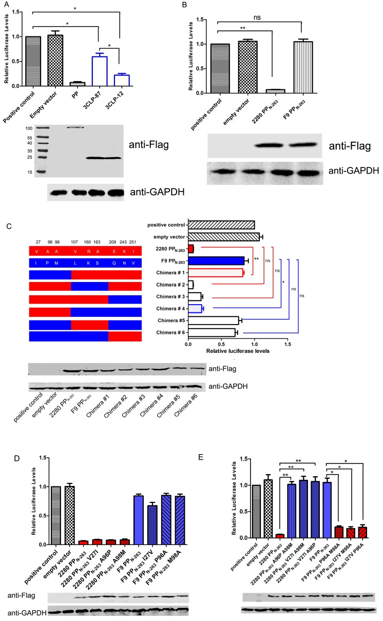
Figure 3.
Identification of the key sites of PP for inhibiting host gene expression. (A,B) CRFK cells were transfected with 0.25 μg of pIFN+33-Luc together with 0.25 μg of pFlag-3CLP-87 and pFlag-3CLP-12 (A); 2280 PPN-263 and F9 PPN-263 (B) or empty vector. At 24 h post-transfection, SeV was inoculated into the cells for 10 h, and then cell lysates were subjected to luciferase assay and Western blot analysis using anti-Flag antibody; (C–E) Luciferase expression from pIFN+33-Luc in cells cotransfected with the indicated chimeric PP genes (C) or single (D) or double mutants (E) of PP in the p3×Flag-CMV vector. The expression of each construct was determined by Western blotting using an anti-Flag antibody. Anti-GAPDH was used as a loading control. Three independent experiments were performed and produced consistent results. The data represented the result of one experiment and were presented as means ± SD. *, p < 0.05; **, p < 0.01; ns, non-significant.

