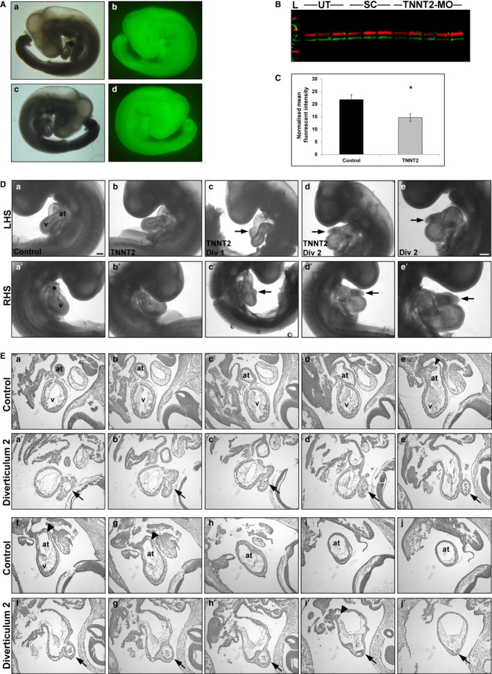
Figure 2.

TNNT2‐MO treatment results in a knockdown of cTNT and external analysis reveals diverticula protruding from the heart wall. (A) Embryos were treated at HH11 with a TNNT2 or standard control fluorescein‐tagged morpholino; only embryos showing a strong fluorescent signal were harvested (at HH19). (b,d) Fluorescent embryo in comparison to brightfield a,c; both left‐ (a,b) and right‐hand side (c,d) shown. (B,C) Western blot of individual hearts treated with either TNNT2‐MO, standard control morpholino or untreated at HH11 results in a significant decrease of cTNT at HH19 (P = 0.016). On (B) the lanes 1–3 are untreated, 4–6 have had standard morpholino applied, and 7–11 are TNNT2‐MO‐treated. (D) External analysis of embryos treated with TNNT2‐MO reveals normal heart development in the majority of embryos (b and b') when compared with the controls (a and a'). However, two embryos presented with diverticula on the heart wall (c and c', d and d'). Diverticulum 2 is shown at higher magnification (e and e'). (E) Serial sectioning through a control (a‐j) and embryo with diverticulum 2 (a'‐j'). Three opening are present between the ventricular chamber into the diverticulum (b', f'and i'). Arrowheads denote the normal atrial septa in controls (f and G), which is reduced in diverticulum 2 heart (i'). *, outflow tract; arrowhead, atrial septum; arrows, diverticulum; at, atrium; LHS, left‐hand side of embryo; RHS, right‐hand side of embryo; v, ventricle; Scale bars: 100 μm.
