Figure 3.

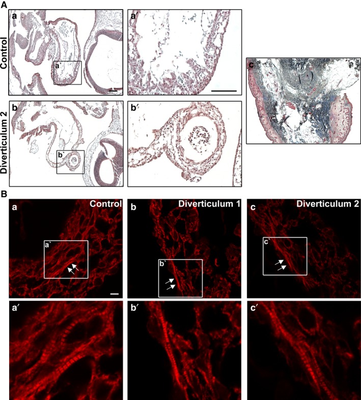
Histological analysis of the diverticula in the TNNT2‐MO‐treated embryos. (A) Masson's Trichrome staining was used to detect collagen that may have been deposited due to fibrosis of the heart. When compared with a control heart (a and a'), the diverticula did not appear to show a noticeable change in collagen deposition that would be indicated by the black/blue staining (b and b'). Chick skin was used as a control for the Trichrome stain (c). Scale bars: (a,a') 300 μm. (B) An anti‐myosin heavy chain antibody was used to detect cardiac muscle in the heart. When compared with the control (a), diverticulum 1 and 2 had normal cardiac muscle appearance, with the presence of mature myofibrils in the myocardial wall (arrows; b and c). a', b' and c' show sarcomeres at higher magnification from images a, b and c, respectively. Scale bar: 80 μm.
