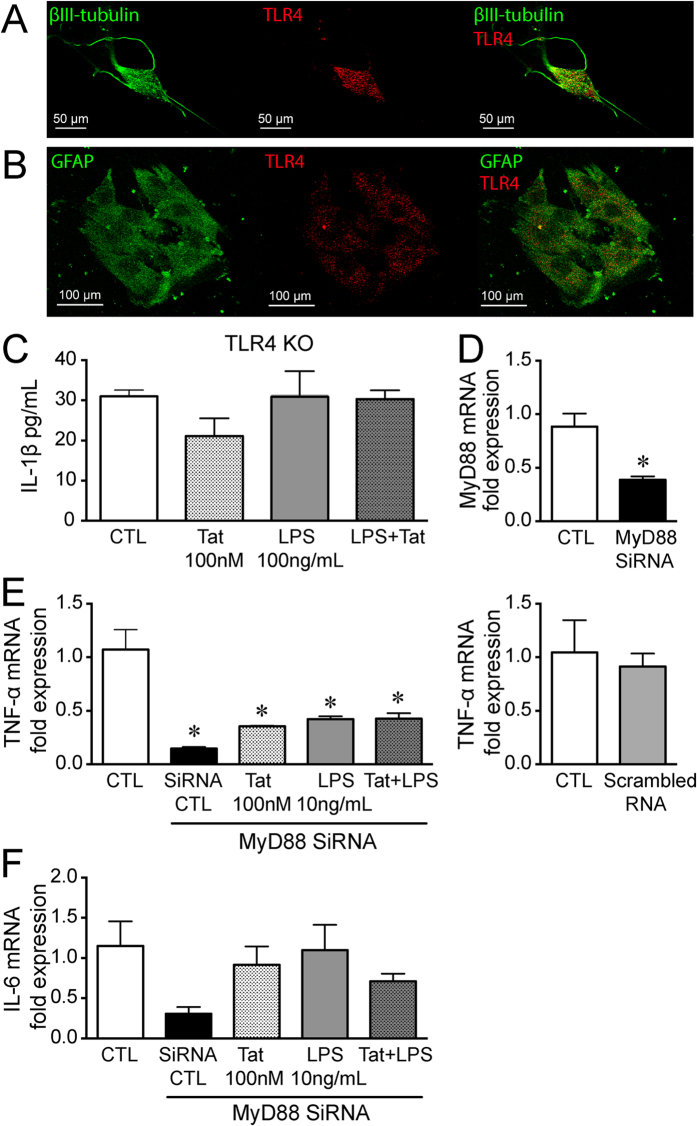
Figure 5. Tat LPS interaction is TLR4 mediated.
(A) Representative confocal microscopic images of enteric β-III tubulin (green) and TLR4 (red) showing the expression of TLR4 on neurons. (B) Representative confocal microscopic images of enteric GFAP (green) and TLR4 (red) showing the expression of TLR4 on enteric glia. (C) Release of IL-1β in neuron-glia co-cultures isolated from TLR4 knockout mice after treatment with 100 nM Tat, 10 ng/mL LPS or Tat+ LPS for 16 h. (D) RT-PCR showing knock down of MyD88 in CRL-2690 enteric glia. (E) Normalized fold mRNA expression of TNF-α and IL-6 (F) in CRL-2690 enteric glia following knock-down of MyD88 or with scrambled RNA and treated with 100 nM Tat, 10 ng/ml LPS or Tat+ LPS. For each set of experiments N = 3, p < 0.05. *vs CTL.

