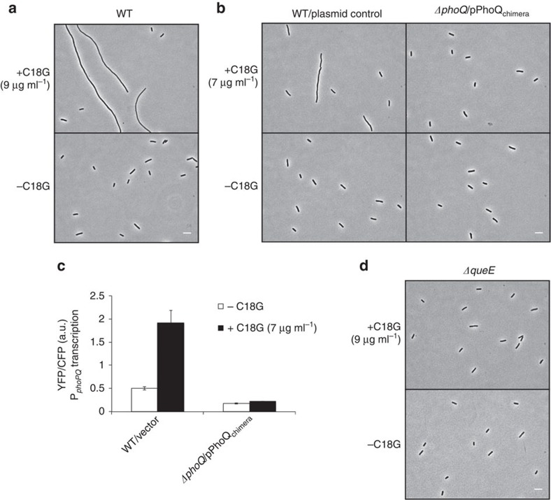
Figure 1. Antimicrobial peptide stimulation of the E. coli PhoQ/PhoP system causes cells to filament.
(a) Phase-contrast micrographs of wild-type (MG1655) cells grown in the presence or absence of C18G. (b) Phase-contrast micrographs of wild-type cells containing control vector (TIM148/pGB2) and ΔphoQ cells containing a plasmid encoding PhoQchimera (TIM229/pLPQ*2) grown in the presence or absence of C18G. (c) Measurement of phoPQ promoter activity (which is regulated by PhoP-P) from a PphoPQ-yfp transcriptional reporter in wild-type cells containing control vector (TIM148/pGB2) and ΔphoQ cells containing a plasmid encoding PhoQchimera (TIM229/pLPQ*2) grown in the presence or absence of C18G. PhoQchimera, which is not stimulated by C18G but has a basal level of PhoQ activity, was used to avoid the high toxicity of C18G in ΔphoQ strains. (See text for details.) Data represent means and s.d.'s from at least three independent experiments. (d) Phase-contrast micrographs of ΔqueE cells (SAM96) grown in the presence or absence of C18G. Cells were grown in minimal medium containing 0.1 mM Mg2+and the indicated concentration of C18G to an OD600=0.2–0.3 and imaged by microscopy. Scale bar, 5 μm.

