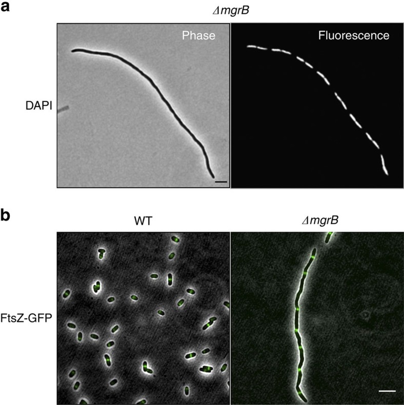
Figure 3. Chromosomal segregation and FtsZ ring formation are not impaired in filamenting cells.
(a) Phase contrast and fluorescence micrographs of ΔmgrB (AML20) cells stained with DAPI. (b) Fluorescence micrographs of wild-type E. coli (EC448) and ΔmgrB (JNC19) cells showing FtsZ-GFP localization. All cultures were grown in minimal medium with no added Mg2+. Scale bar, 5 μm.

