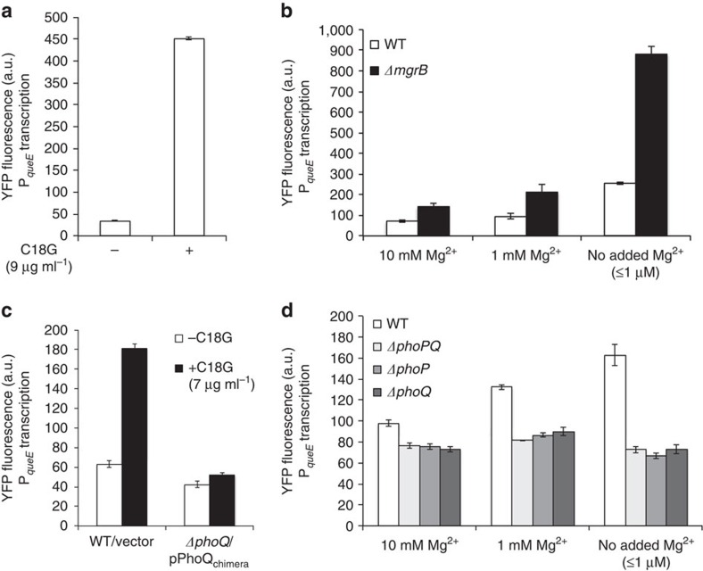
Figure 4. The PhoQ/PhoP system regulates queE transcription.
(a) YFP fluorescence of a queE-yfp operon fusion measured in wild-type cells (SAM54) growing in the presence (+) or absence (−) of C18G (9 μg ml−1) in minimal medium containing 0.1 mM Mg2+. Data represent the means and ranges of two independent cultures. (b) YFP fluorescence measured from a queE-yfp operon fusion in wild-type (SAM54) and ΔmgrB (SAM55) strains in minimal medium at the indicated Mg2+ concentrations. Data represent means and s.d.'s from at least three independent experiments. (c) YFP fluorescence of a queE-yfp operon fusion measured in wild-type cells containing a control plasmid (SAM54/pGB2) and ΔphoQ cells containing a plasmid encoding PhoQchimera (SAM60/pLPQ*2) grown in the presence (+) or absence (−) of C18G (7 μg ml−1) in minimal medium containing 0.1 mM Mg2+. Data represent the means and s.d.'s from at least three independent experiments. The PhoQchimera protein, which is not stimulated by C18G but has a basal level of PhoQ activity, was used to avoid problems due to the high toxicity of C18G in ΔphoQ strains. (d) YFP fluorescence measured from a queE-yfp operon fusion in wild-type (SAM54), ΔphoPQ (SAM141), ΔphoP (SAM142) and ΔphoQ (SAM60) strains in minimal medium at the indicated Mg2+ concentration. Data represent the means and ranges of two independent cultures.

