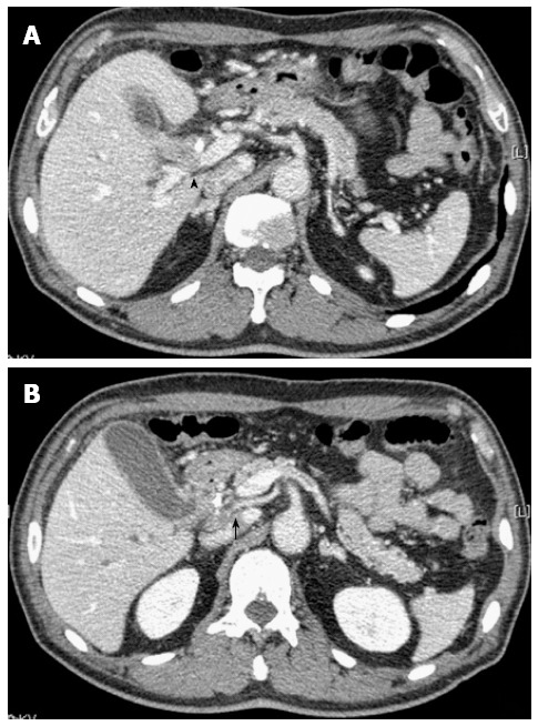
Figure 2.

Abdominal computed tomography images at two different levels are shown. A: Computed tomography (CT) showed a low-density mass measuring 18 mm in diameter located in the middle bile duct adjacent to the portal vein (arrowhead). An endoscopic nasobiliary drainage tube was observed in the bile duct; B: CT showed a low-density mass involving the right hepatic artery from the superior mesenteric artery in the middle bile duct (arrow).
