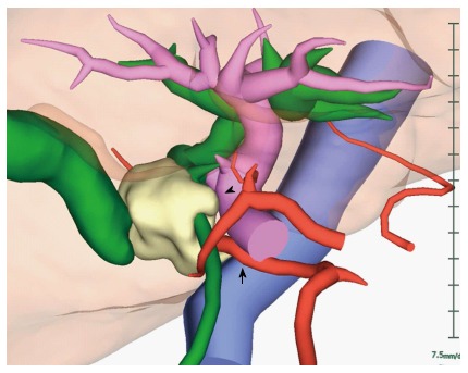
Figure 3.

3D reconstructed computed tomography fused with an magnetic resonance cholangiopancreatography image. The tumor was located in the middle bile duct. The tumor was suspected to have invaded the portal vein (arrowhead) and the right hepatic artery from the superior mesenteric artery (arrow).
