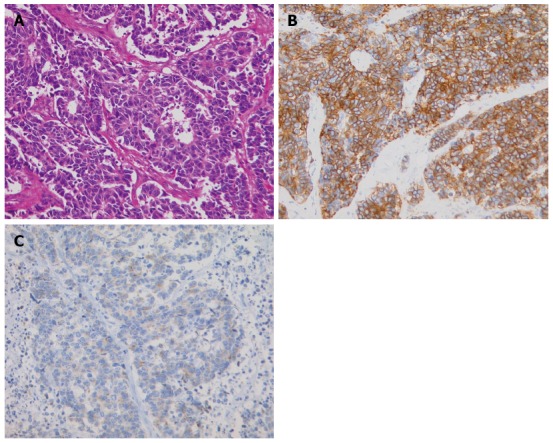
Figure 4.

Histopathologic appearance of the neuroendocrine carcinoma. A: The cells were round or oval, hyperchromatic, and had an increased nucleus-to-cytoplasm ratio (hematoxylin-eosin staining, magnification × 200); B: Immunohistochemically, tumor cells were diffusely positive for CD56, a membrane protein usually present in neuroendocrine cells; C: Immunohistochemically, tumor cells were positive for synaptophysin, which is typically expressed on the surface of neurons or endothelial cells.
