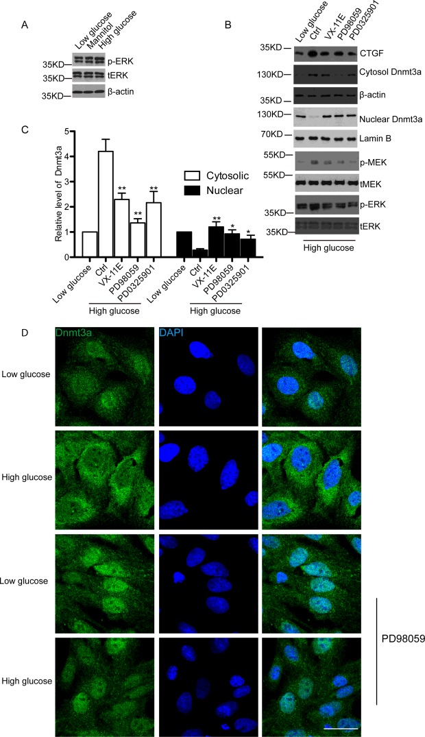
Figure 4. ERK/MEK signalling pathway contributes to cytoplasmic translocation of Dnmt3a in high glucose-treated hMSCs.
(A) Western blot detected the pERK and total ERK (tERK) in low glucose, mannitol or high glucose-treated hMSCs. β-Actin was used as a loading control. n=3 and representative image was shown. (B) Western blot analysis of CTGF, pMEK, total MEK (tMEK), pERK, tERK, cytoplasmic and nuclear Dnmt3a protein levels at 24 h post high glucose treatment followed by 12 h treatment of control, 0.5 μM ERK inhibitor VX-11E, 50 μM MEK inhibitor PD98059 or 0.5 μM MEK inhibitor PD0325901. β-Actin was used as the internal control for cytoplasmic portion and Lamin B was used as the internal control of nuclear portion. n=3 and representative image was shown. (C) Quantitative analysis of (B) by ImageJ software based on three independent experiments. *P<0.05. **P<0.01. (D) Immunofluorescence staining of Dnmt3a (green) under low glucose, mannitol or high glucose conditions for 24 h with or without 50 uM PD98059. DAPI (blue) was used to stain the nucleus. At least 10 microscopic fields were assessed in each experiment. Images are representative of three independent experiments. Scale bar, 10 μm.

