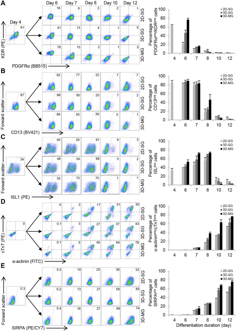
Figure 5. Simulated microgravity and 3D culture promote the induction of cardiac progenitors and CM differentiation.
Flow cytometry analysis of cells exposed to microgravity (3D-MG) and standard gravity (2D-SG and 3D-SG) at various time points for their expression of markers associated with (1) cardiac mesoderm, (A) KDR/PDGFRα and (B) CD13; (2) cardiac progenitors, (C) ISL1; and (3) cardiomyocytes, (D) SIRPA and (E) cTnT/α-actinin. Data are presented as representative flow cytometry analysis and summary based on mean ± SD of 3 biological replicates.

