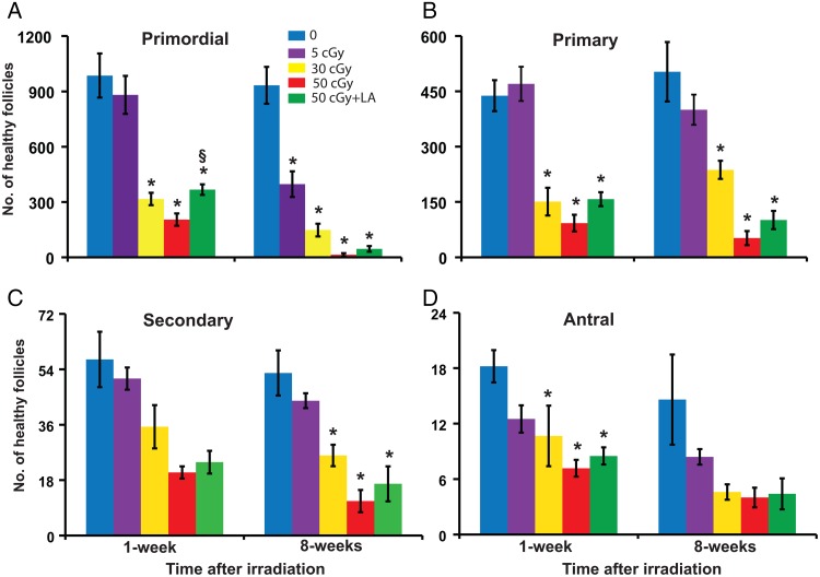
Figure 1.
Charged iron particles deplete ovarian follicles. Mice in this and subsequent figures were fed normal diet or diet supplemented with 150 mg/kg ALA from 1 week before irradiation with the indicated doses of charged iron particles until euthanasia. Graphs show the mean ± SEM of healthy follicles of the indicated stages of development per ovary. (A) Primordial follicles, P < 0.001, effect of group by ANOVA at both time points. (B) Primary follicles, P < 0.001, effect of group by ANOVA at both time points. (C) Secondary follicles, P ≤ 0.001, effect of group by ANOVA at both time points. (D) Antral follicles P ≤ 0.011, effect of group by ANOVA at both time points. *P < 0.05, compared with 0 cGy control; §P < 0.05, 50 cGY + ALA compared with 50 cGy. n = 5–6/group.

