Figure 4.
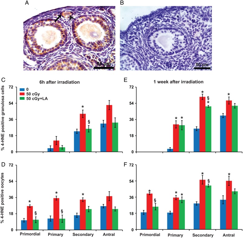
Charged iron particles increase lipid peroxidation in ovarian follicles. (A) Representative image of 4-HNE localization in granulosa cells (black arrow) and oocyte (arrowhead) of follicles. (B) Negative control with primary antibody replaced by nonimmune IgG. Graphs show the means ± SEM of percentages of follicles with 4-HNE positive granulosa cells or oocytes. (C) Six hours after irradiation, percentages of follicles with 4-HNE positive granulosa cells varied significantly among groups for secondary follicles (P < 0.05, Kruskal–Wallis test) and approached significance for antral follicles (P = 0.08). (D) Percentages of follicles with 4-HNE positive oocytes varied significantly among groups for primordial, primary and secondary follicles (P < 0.05, Kruskall–Wallis test) and approached significance for antral follicles (P = 0.06). (E) One week after irradiation, percentages of primary, secondary and antral follicles with 4-HNE positive granulosa cells varied significantly among groups (P < 0.05, Kruskal–Wallis test). (F) At 1 week, percentages of follicles with 4-HNE positive oocytes varied significantly among groups for primordial, primary, secondary and antral follicles (P < 0.05, Kruskal–Wallis test). *P < 0.05 versus 0 cGy control by Mann–Whitney test; §P < 0.05, 50 cGY + ALA versus 50 cGy by Mann–Whitney test. n = 4 mice/group.

