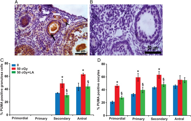
Figure 7.
Induction of apoptosis in ovarian follicles by charged iron particles involves PUMA. (A) Representative image of PUMA localization in granulosa cells (black arrow) and oocytes (arrowhead) of follicles. (B) Negative control image with primary antibody replaced by nonimmune IgG. Graphs show the means ± SEM of percentages of follicles with PUMA-positive granulosa cells or oocytes at 6 h after irradiation. (C) Percentages of secondary and antral follicles with PUMA-positive granulosa cells varied significantly among groups (P < 0.05, Kruskal–Wallis test). (D) Percentages of primordial, primary, and secondary follicles with PUMA-positive oocytes varied significantly among groups (P < 0.05, Kruskal–Wallis test). *P < 0.05 versus 0 cGy control by Mann–Whitney test; §P < 0.05, 50 cGY + ALA versus 50 cGy by Mann–Whitney test. n = 4 mice/group.

