Fig. 1.

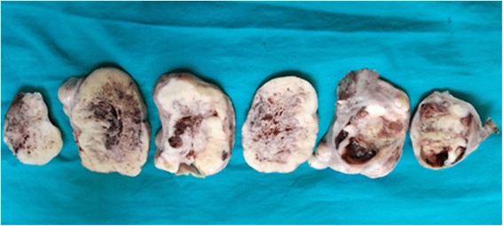
Left salpingo-oophorectomy specimen showing a 15x10x5 cm ovarian mass with gray white solid fleshy areas, interspersed with areas of necrosis, hemorrhage, and cystic spaces

Left salpingo-oophorectomy specimen showing a 15x10x5 cm ovarian mass with gray white solid fleshy areas, interspersed with areas of necrosis, hemorrhage, and cystic spaces