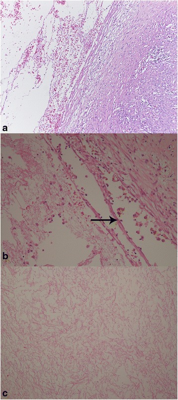
Fig. 2.

R1: Microscopic examination revealed that the cystic spaces showed no epithelial lining (a: Hematoxylin-eosin x 100). Note the presence of macrophages filled with hemosiderin in the wall of these spaces (b: Hematoxylin-eosin, Arrow x 200) and fibrin with pink network aspect in the cavity of cystic space (c: Hematoxylin-eosin, x 200)
