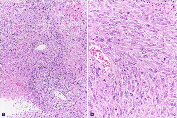
Fig. 3.

The solid component of the neoplasm contained large foci of coagulative tumor cell necrosis and spindle cells, with moderate cytoplasm, arranged predominantly in fascicles or a storiform pattern, mimicking mesenchymal malignancy (a: Hematoxylin-eosin x 100). In other smaller areas, the neoplastic elements showed abundant eosinophilic cytoplasm. The nuclei were atypical and vesicular, with evident nucleoli. Mitoses were frequent and sometimes atypical (b: Hematoxylin-eosin x 400)
