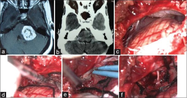
Figure 3.

Preoperative axial T1-weighted contrast magnetic resonance imaging showing left cerebellopontine (CP) angle tumor (a), postoperative contrast computed tomography scan reveal complete removal of tumor (b), Intraoperative video telescopic operating monitor view of CP angle (c), tumor debulking (d), separation from brain stem (e) and after tumor removal and anatomical preservation of facial nerve (f)
