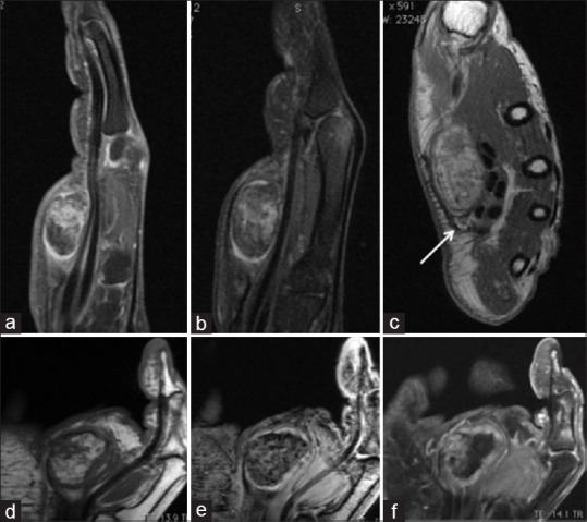
Figure 2.

Hand magnetic resonance image demonstrating the well-encapsulated fatty-fibrous tumor of the median nerve. Sagittal T1-weighted with gadolinium (a) and fat suppression (b) demonstrating the relationship with the palmar tendons. Axial T1-weighted (c) demonstrating the heterogeneous mass attached to the median nerve (arrow). Coronal T1 (d) demonstrates similar fat signal of the hipotenar region complemented with coronal T2 gradient echo (e) and T1 with gadolinium (f)
