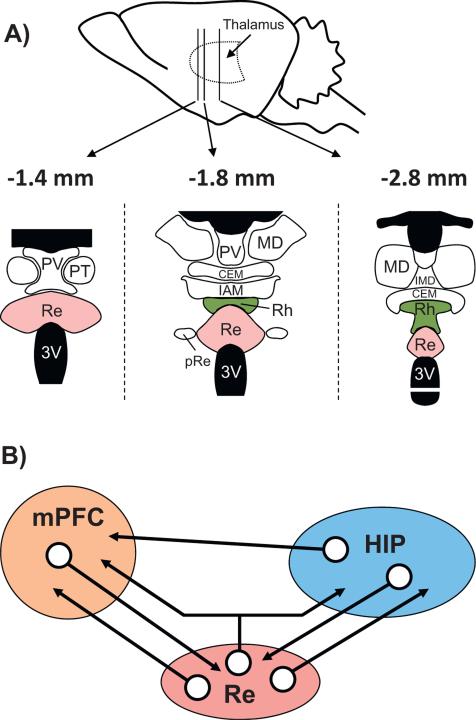
Fig. 1.
Neuroanatomical organization of the midline thalamus (A) and connectivity diagram between the prefrontal cortex, the reuniens nucleus and the hippocampus (B). A: Nuclei of the ventral midline thalamus, with particular focus on the reuniens (Re) and rhomboid (Rh) nuclei at 3 anterior-posterior levels (the most caudal is on the left). Abbreviations: CEM, central medial nucleus; IAM, interanteromedial nucleus; IMD, interomediodorsal nucleus; MD, mediodorsal nucleus; pRe, perireuniens nuclei; PT, paratenial nucleus; PV, paraventricular nucleus; Re: reuniens nucleus; Rh, rhomboid nucleus. B: Schematic representation of the organization of the network connectivity within a system formed by the reuniens nucleus (Re), which is the largest of the ventral midline thalamus, the hippocampus (HIP) and the medial prefrontal cortex (mPFC). It is noteworthy that whereas the HIP has pronounced projections to the mPFC, there are no direct return ones from the mPFC to the hippocampus. The Re has dense projections to both the mPFC and the HIP (see also Fig. 2), and a small proportion of neurons of the Re (between 3 and 6%) even send axon collaterals to both structures, as described very recently (Hoover and Vertes, 2012). Finally, the mPFC and the HIP have dense projections to the Re/Rh. This organization places the Re, and perhaps more generally the ventral midline thalamus (which also encompasses the rhomboid and perireuniens nuclei), in a pivotal position to influence prefrontal cortical and hippocampal functions, and perhaps even in a more specific way functions which depend on information exchange between or coordination of these two structures.

