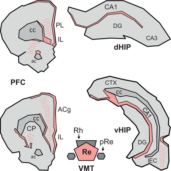
Fig. 2.
Terminal projection fields of the reuniens nucleus in the prefrontal cortex and the hippocampus of the Rat. Illustration of the location and extent of the regions of densest (filled) and weaker (hatched) fiber staining at two A-P levels of the medial prefrontal cortex (mPFC) and in the dorsal and ventral hippocampus (dHIP and vHIP, respectively) produced by an injection of the anterograde anatomical tracer Phaseolus vulgaris leucoagglutinin into the Re. The drawings were made according to the darkfield microphotographs (Figs. 5 and 6) and the schematic representations (Fig. 3) shown in Vertes et al. (2006). Abbreviations: ac, anterior commissure; ACg, anterior cingulate cortex; cc, corpus callosum; CP, caudate putamen; DG, dentate gyrus; IL, infralimbic cortex; LEC, lateral entorhinal cortex; pRe, perireuniens nucleus; PL, prelimbic cortex; Re, reuniens nucleus; Rh, rhomboid nucleus.

