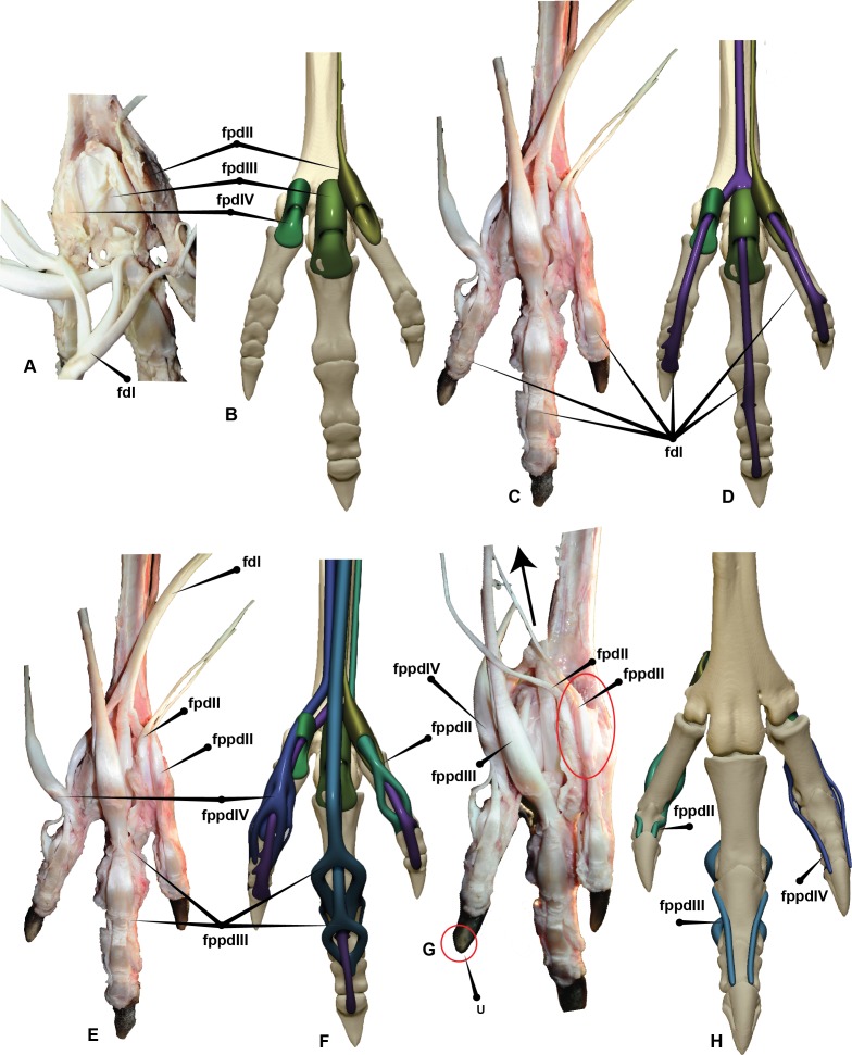
Figure 7. Dromaius pes with intermediate, superficial and deep flexors.
Plantar view displaying: (A) Extension of superficial flexors for each corresponding digit that housed the deep and intermediate flexors. (B) Corresponding digital render of superficial flexors. (C) Ventral dissection identifying the superficial flexors (cartilage housing for the tendons) and the flexor digitorum longus. (D) Corresponding digital rendering of dissection. (E) Ventral dissection identifying intermediate, superficial and deep tendons. (F) Corresponding digital rendering of dissection. (G) Close up of the digit II’s superficial flexor. (H) Cranial view of the intermediate flexor attachments. Abbreviations: M. flexor digitorum longus (fdl); M. flexor hallucis brevis (fhb); M. flexor hallucis longus (fhl); M. extensor digitorum longus (edl); M. flexor perforatus digiti II (fpdII); M. flexor perforans et perforatus digiti II (fppdll); flexor perforates digiti III (fpdIII); M. flexor perforans et perforatus digiti III (fppdIII); flexor perforatus digiti IV (fpdIV).

