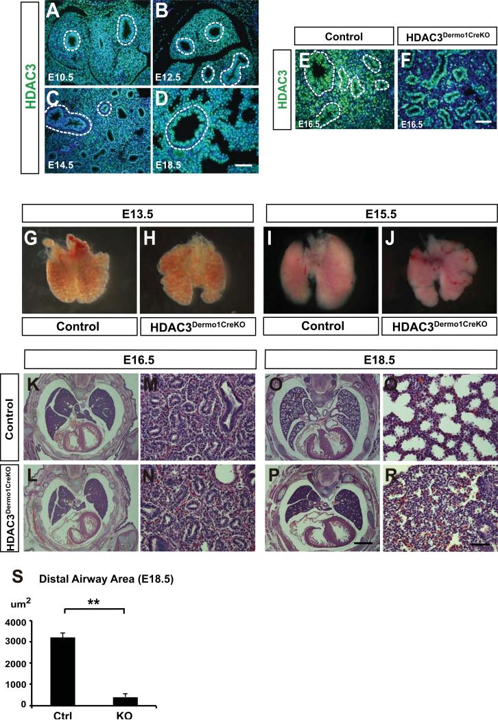
Figure 1. Loss of HDAC3 in the lung mesenchyme leads to hypoplasia and sacculation defects.
(A-D) HDAC3 is broadly expressed in both lung epithelium and mesenchyme from E10.5 to E18.5. Dotted lines mark the boundary between lung epithelium and mesenchyme. (E-F) HDAC3 is efficiently deleted using the Dermo1cre lines as noted by loss of HDAC3 expression in the developing lung mesenchymal cells using immunostaining. Dotted lines mark the boundary between lung epithelium and mesenchyme. (G-H) At E13.5, the Hdac3Dermo1creKO mutants show no obvious defects in lung morphology. (I-J) At E15.5, Hdac3Dermo1creKO lungs exhibit a reduced size shown by the whole-mount pictures. (K-N) H&E staining show that the Hdac3Dermo1creKO lungs exhibit normal epithelial branching. (O-S) The Hdac3Dermo1creKO mutants display disrupted lung sacculation at E18.5 as exhibited by reduced distal airspace area.
Two tail student's t test: **p<0.01. n=3. Q-PCR data are represented as mean ± SD. Scale bars: D, F and R=50μm; P=1mm.

