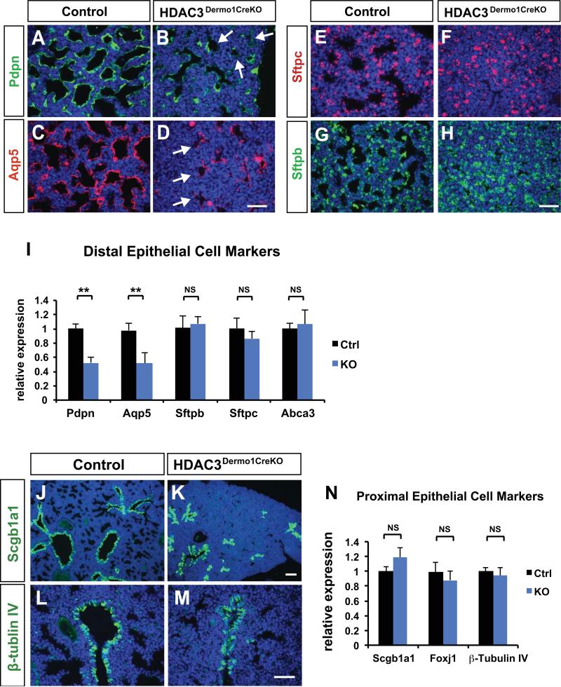
Figure 2. Loss of HDAC3 in the developing lung mesenchyme results in a loss of AT1 cell differentiation.
(A-D) Immunostaining for type I alveolar epithelial cell makers Pdpn and Aqp5 is significantly decreased in the Hdac3Dermo1creKO mutant lungs at E18.5 (arrows). (E-H) Expression of the alveolar type 2 cell (AT2) marker Sftpc and Sftpb are unchanged in the Hdac3Dermo1creKO mutants as revealed by immunostaining. (I) Q-PCR results confirm the loss of Pdpn and Aqp5 expression in mutant lungs at E18.5, and normal expression of AT2 cell specific makers at E18.5. (J-M) Expression of the secretory epithelium lineage marker Scgb1a1 and markers of the ciliated epithelial lineage TubbIV and Foxj1 are unchanged in the Hdac3Dermo1creKO mutants as revealed by immunostaining. (N) Q-PCR results also indicate normal expression of these cell specific makers at E18.5.
Ctrl=Control; KO= Hdac3Dermo1creKO lungs. Two tail student's t test: **p<0.01; NS=Not Significant. n=6. Q-PCR data are represented as mean ± SD.
Scale bars: D, H and M=50μm; K=100μm.

